PARIETAL EYE “PHOTORECEPTIVE” THIRD EYE ?LIGHT INFORMATION “CRYSTAL”
THE PINEAL GLAND THE VISION THE EYE ~ INTUITION
?
The results of various scientific research in evolutionary biology, comparative neuroanatomy and neurophysiology, have explained the phylogeny of the pineal gland in different vertebrate species. From the point of view of biological evolution, the pineal gland represents a kind of atrophied photoreceptor. In the epithalamus of some species of amphibians and reptiles, it is linked to a light-sensing organ, known as the parietal eye, which is also called the pineal eye or third eye.[8]
René Descartes believed the pineal gland to be the “principal seat of the soul.” Academic philosophy among his contemporaries considered the pineal gland as a neuroanatomical structure without special metaphysical qualities; science studied it as one endocrine gland among many.
The pineal gland is a midline brain structure that is unpaired. It takes its name from its pine-cone shape.[1][9] The gland is reddish-gray and about the size of a grain of rice (5–8 mm) in humans. The pineal gland, also called the pineal body, is part of the epithalamus, and lies between the laterally positioned thalamic bodies and behind the habenular commissure. It is located in the quadrigeminal cistern near to the corpora quadrigemina.[10] It is also located behind the third ventricle and is bathed in cerebrospinal fluid supplied through a small pineal recess of the third ventricle which projects into the stalk of the gland.[11]
Blood supply
Unlike most of the mammalian brain, the pineal gland is not isolated from the body by the blood–brain barrier system;[12] it has profuse blood flow, second only to the kidney,[13] supplied from the choroidal branches of the posterior cerebral artery.
Nerve supply
The pineal gland receives a sympathetic innervation from the superior cervical ganglion. A parasympathetic innervation from the pterygopalatine and otic ganglia is also present.[14] Further, some nerve fibers penetrate into the pineal gland via the pineal stalk (central innervation). Also, neurons in the trigeminal ganglion innervate the gland with nerve fibers containing the neuropeptide PACAP.


The pineal body consists in humans of a lobular parenchyma of pinealocytes surrounded by connective tissue spaces. The gland’s surface is covered by a pial capsule.
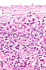
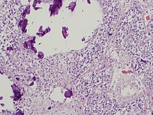
The pineal gland consists mainly of pinealocytes, but four other cell types have been identified. As it is quite cellular (in relation to the cortex and white matter), it may be mistaken for a neoplasm.[15]
FUNCTION
The primary function of the pineal gland is to produce melatonin. Melatonin has various functions in the central nervous system, the most important of which is to help modulate sleep patterns. Melatonin production is stimulated by darkness and inhibited by light.[27][28] Light sensitive nerve cells in the retina detect light and send this signal to the suprachiasmatic nucleus (SCN), synchronizing the SCN to the day-night cycle. Nerve fibers then relay the daylight information from the SCN to the paraventricular nuclei (PVN), then to the spinal cord and via the sympathetic system to superior cervical ganglia(SCG), and from there into the pineal gland.
The compound pinoline is also claimed to be produced in the pineal gland; it is one of the beta-carbolines.[29] This claim is subject to some controversy.
Regulation of the pituitary gland
Studies on rodents suggest that the pineal gland influences the pituitary gland‘s secretion of the sex hormones, follicle-stimulating hormone (FSH), and luteinizing hormone (LH). Pinealectomy performed on rodents produced no change in pituitary weight, but caused an increase in the concentration of FSH and LH within the gland.[30] Administration of melatonindid not return the concentrations of FSH to normal levels, suggesting that the pineal gland influences pituitary gland secretion of FSH and LH through an undescribed transmitting molecule.[30]
The pineal gland contains receptors for the regulatory neuropeptide, endothelin-1,[31] which, when injected in picomolarquantities into the lateral cerebral ventricle, causes a calcium-mediated increase in pineal glucose metabolism.[32]
*Material Provided By: WIKIPEDIA


